Project Overview
Detection of COVID-19 Positive Cases Using Deep Learning leverages AI to classify chest X-ray images into categories: COVID-positive, lung opacity, normal, and viral pneumonia. This project aims to support healthcare providers with quick, accurate preliminary diagnoses, easing the strain on medical resources during high-demand periods.
The link to the code is here on GitHub: GitHub and dataset link of kaggle: Dataset
This project applies deep learning techniques to detect COVID-19 positive cases from chest X-ray images. By categorizing images as COVID-positive, lung opacity, normal, or viral pneumonia, this model aims to assist healthcare providers in identifying cases quickly and accurately. It aims to assist healthcare providers with quick, accurate preliminary diagnoses, alleviating pressure on medical resources, especially during high-demand periods like pandemics.
The dataset is sourced from COVID-19 Radiography Database on Kaggle, consisting of X-ray images classified into COVID, Normal, Lung Opacity, and Viral Pneumonia categories.
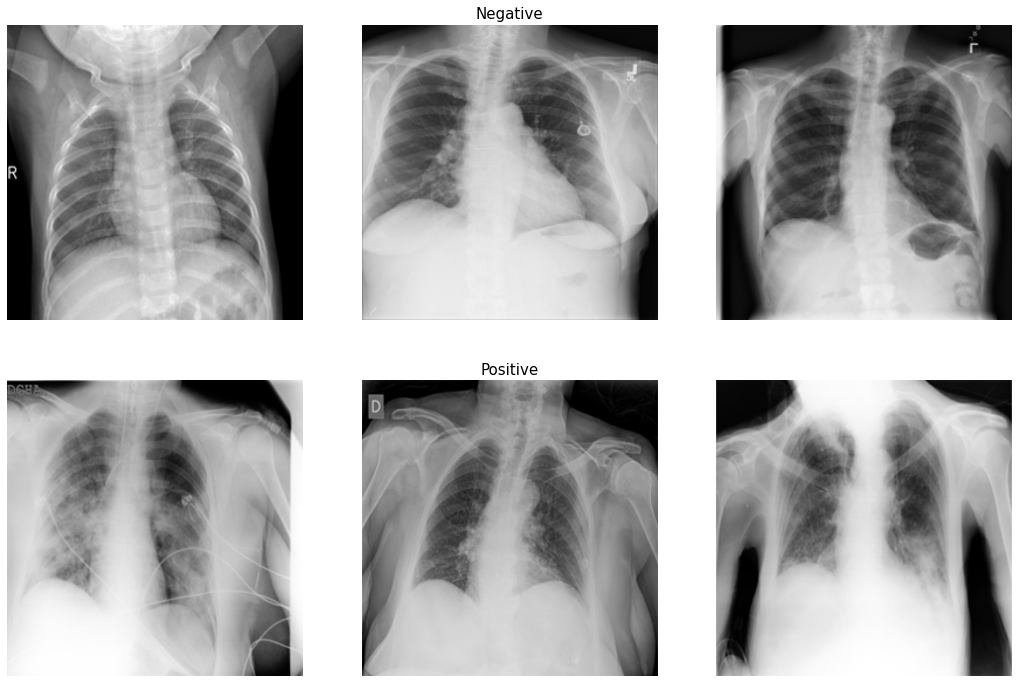
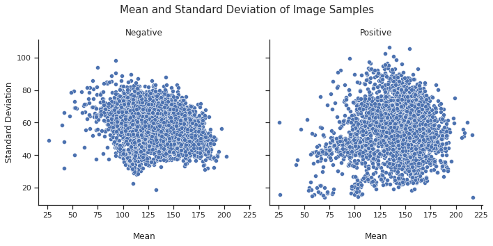
Exploratory Data Analysis (EDA) EDA involves visualizing the data distribution for different classes. Count plots show the number of COVID-positive vs. negative cases, and sample images help understand image characteristics, which is essential for effective model
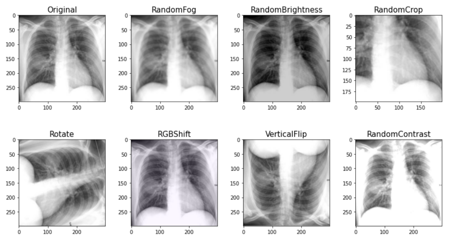
- Image Preprocessing and
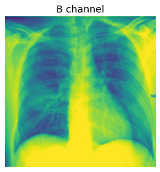
Augmentation Image preprocessingincludes resizing and converting images to grayscale, with additional augmentations like rotation, brightness adjustment, and flipping. These steps improve model generalization and simulate varied conditions within the dataset. Ben Graham's MethodUsing Ben Graham's Method, images are converted to grayscale and smoothed with aGaussian blur. This step reduces noise and highlights important structures in X-ray images, enabling the model to focus on relevant patterns associated with COVID-19.- Model Building A
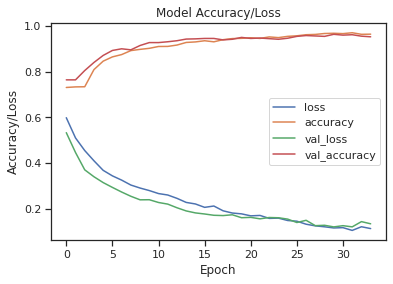
Convolutional Neural Network (CNN)model is designed with layers for feature extraction and classification. The CNN leverages imagepatternsto classify X-rays, focusing on detecting features related to COVID-19. - Model Training The model is trained on the dataset with validation monitoring to
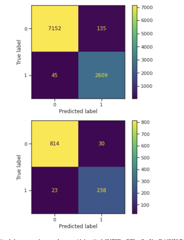
prevent overfitting. Techniques likeearly stopping and dropout regularizationare used to enhance model performance, providing a balance between training and validation accuracy. - Model Evaluation After training, the model’s performance is evaluated using metrics such as accuracy, recall, and F1 score on the test set.
Confusion matricesgive insights into classification accuracy across different classes. Grad-CAM VisualizationGrad-CAM is used to generate heatmaps for COVID-positive and negative cases, highlighting areas in the X-rays where the model focuses to make its predictions. This step providesinterpretabilityby showing which regions influenced the model’s decision.
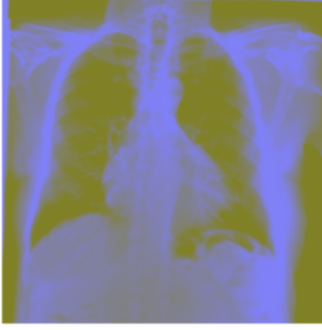

Grad-CAM (Gradient-weighted Class Activation Mapping) is a powerful technique for visualizing and interpreting the inner workings of Convolutional Neural Networks (CNNs), especially in image classification tasks. Grad-CAM is used to highlight the areas of an image that the model deems most important for its prediction, offering insights into how the model is making its decisions. The technique is especially beneficial for complex tasks, such as medical image classification (e.g., detecting diseases in chest X-rays), where understanding the model’s reasoning is critical.
Grad-CAM uses the gradients of the class score (the probability for the predicted class) with respect to the feature maps of the last convolutional layer. These gradients represent how sensitive the model is to changes in each feature map for a given image.
Forward Pass: The image is passed through the model to obtain a class prediction.
The gradients of the predicted class with respect to the last convolutional layer's feature maps are calculated.
Pooling the Gradients: The gradients are globally pooled (usually averaged) to obtain weightsthat describe the importance of each feature map.
Weighting the Feature Maps: Each feature map in the last convolutional layer is multiplied by its corresponding gradient weight.
Grad-CAM increases the transparency of the model's inner workings, which is crucial in medical AI applications. This interpretability is especially important in high-stakes domains like healthcare, where a model’s output can directly influence patient care.
Generating the Heatmap The weighted feature maps are summed and passed through a ReLU activation(to remove negative values), generating a class-discriminative heatmap.
Superimposition: The heatmap is then resized to match the dimensions of the input image and is overlaid on the original image to highlight the regions most influential for the model's decision.
By integrating Grad-CAM, I was able to provide a deeper level of interpretability and validation for the model, making it not only more reliable but also understandable in a critical field like healthcare.
Also. If you need someone to talk to, please reach out. I'm always here and happy to talk.